Imaging
Diagnostic Testing
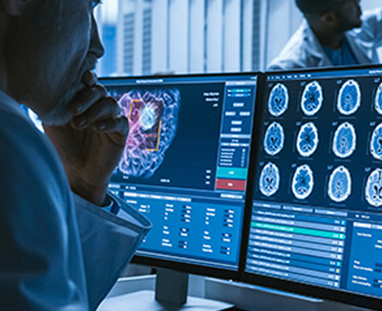
Saint Mary’s diagnostic tools provide the highest quality of care and improved patient outcomes. We offer a wide range of services and our Picture Archiving Communication Systems (PACS) platform allows for rapid access to images for radiologists and clinicians. Please fill out the appointment request form to select your desired time, location, and imaging service needed, and a representative will contact you to complete the scheduling process. Please note, no appointments or request forms are needed for standard X-ray. Patients are welcome to walk-in at any of our locations during operating hours to receive their X-ray.
For further information, contact us at 775-770-3187
Patient Reviews
Bone Density Scanning (DEXA)
DEXA is the preferred technique for measuring bone mineral density (BMD). DEXA scanning focuses on two main areas of the skeleton, the hip and spine. Although osteoporosis involves the whole body, measurements of BMD can be predictive of fractures.
MRI
Our new state of the art 3T Vida scanner with BioMatrix technology automatically adjusts to you to deliver an MRI that is precise, comfortable, and fast.
CT
Computed Tomography (CT or CAT scan) combines a series of X-ray views taken from many different angles and computer processing to create cross-sectional images of the bones and soft tissues inside the body. Saint Mary’s now offers a 64 slice CT scanner. The GE 660 is the latest in its class and is twice as fast as the typical 64 slice scanner. For precise guidance when performing biopsies and drainages we offer CT guided interventions.
Interventional Radiology
Ultrasound
Cardiovascular Ultrasound
In our vascular ultrasound area we obtain real-time images of arteries and veins in the body to assess blood flow and abnormalities. We can evaluate blood flow to the arms, legs, brain, and kidneys. We also perform detailed mapping of the veins contributing to varicosities and lower extremity wounds.
We offer state of the art cardiac ultrasound technology to support Saint Mary’s Structural Heart program. With this latest technology we can capture the anatomy and structure of the heart prior to cardiac interventions including TAVR and Mitraclip.
Nuclear Medicine
At our downtown campus we have two GE discovery dual head gamma cameras with advanced software applications that are much faster and provide significantly improved image resolution. At the Center For Health, a dedicated triple headed gamma camera for cardiac imaging has been installed.
PET
Our combined PET/CT scans provide images that pinpoint the anatomic location of abnormal metabolic activity within the body. The combined scans have been shown to provide more accurate diagnoses than the two scans performed separately as in PET/CT fusion.
X-RAY
Women’s Imaging Services
Mammography
Automated Breast Ultrasound (ABUS)
Saint Mary’s now offers the area’s first FDA approved whole breast ultrasound system for women who have dense breast tissue. The Invenia ABUS can provide additional information when used in conjunction with mammography.
ABUS is covered by most insurances
SAVI SCOUT®
In seeking a more compassionate and precise approach to breast cancer tumor localization, Saint Mary’s Health Network has adopted the SAVI SCOUT® wire-free radar localization system for breast conserving surgeries. Previously surgeons relied on placement of a wire into the tumor adding additional time, inconvience, and discomfort on the day of surgery. SAVI SCOUT® is a new efficient and precise approach to localization and surgical guidance and helps surgeons remove cancerous tissue with greater confidence.
Prior to SCOUT, the most common approach for localizing breast tumors was wire localization. With wire localization, a radiologist would place a thin, hooked wire through the skin to the tumor location. The surgeon would then use the wire to locate the tumor. This procedure would be done on the day of surgery and because the time between the wire placement and surgery can be several hours, this added procedure can be the cause of added worry and discomfort.
The SCOUT® wire-free radar localization system is a clinically proven, FDA cleared, zero-radiation approach to targeting tumors and breast lesions. SCOUT uses a unique radar signal to detect a reflector, the size of a grain of rice, that can be placed at the tumor site at any time during the course of treatment and at the patients’ convenience. During the procedure, the surgeon scans the breast using the SCOUT guide to precisely and efficiently locate the reflector to within 1mm of accuracy.
The ability to precisely locate tumors increases the probability of complete cancer removal and reduces the likelihood of needing follow-up surgeries – a huge advantage for early-state breast cancer patients. In addition, the ability to strategically plan the incision may result in better cosmetic outcomes.
SCOUT is also uniquely suited for marking tumors and lymph nodes prior to neoadjuvant therapy because the SCOUT reflector does not interfere with imaging studies that are needed after treatment to see if the tumor has shrunk.

Locations
Patients now have access to the latest in imaging technology and benefit from an outpatient pricing structure that is more affordable and competitively priced compared to local diagnostic centers. To schedule an appointment, call 775.770.3187.
Saint Mary’s Health Network
235 West Sixth Street
Monday – Friday 7 a.m. to 5 p.m.
775-770-3187
Saint Mary’s Center for Health
645 N. Arlington Ave., Suite 250
Monday – Friday 7 a.m. to 5 p.m.
775-770-3187
South Reno
18653 Wedge Pkwy
Monday – Friday 7 a.m. to 5 p.m.
775-770-3187
North Valleys
280 Vista Knoll Parkway,
Suite 106. Reno, NV 89506
Monday – Friday 7 a.m. to 5 p.m.
775-770-3187
Northwest
6255 Sharlands Ave.
Monday – Friday 7 a.m. to 5 p.m.
X-Rays Daily 7 a.m. to 5 p.m.
775-770-3187
Spanish Springs
X-ray services only (Daily)
5070 Ion Drive
Monday – Friday 7 a.m. to 5 p.m.
775-770-3187

Featured Services

Behavioral Health

Emergency Services

Lab Services
Primary Care


